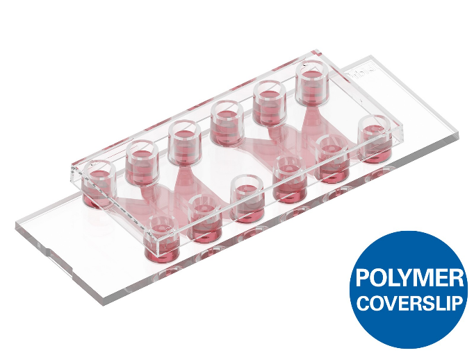
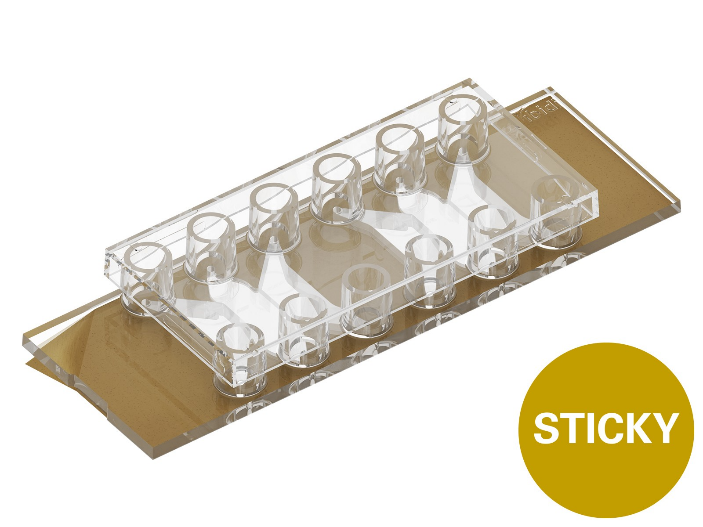
sticky-Slide Tissue Engineering: Highly versatile: sticky side can be mounted on a variety of coverslips or other bottom materials
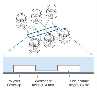
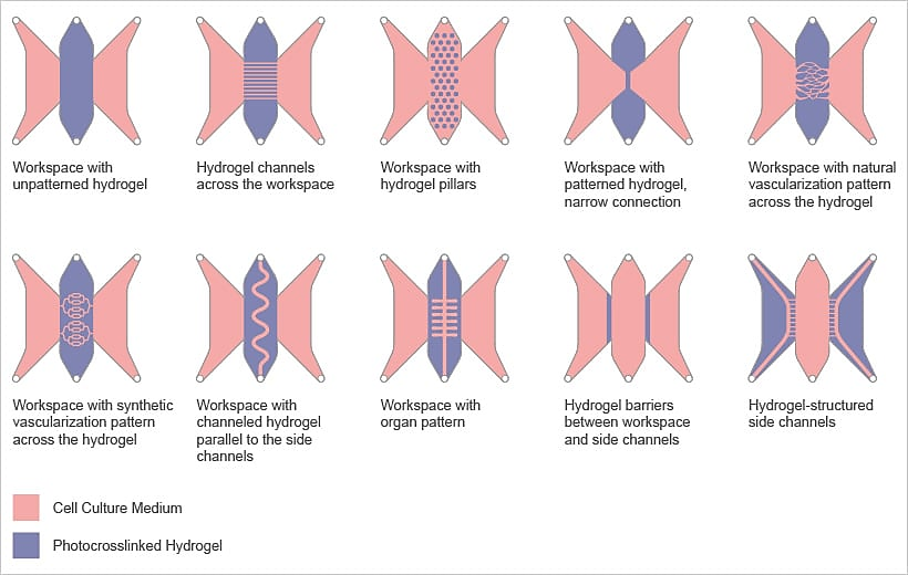
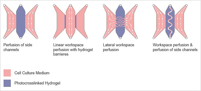
The central workspace and side channels can be structured using photocrosslinkable hydrogels, defining the locations for the cell/tissue model and fluidic access.
Applications
Tissue and Organ-on-a-Chip Models
Custom 3D structures: Fabricate microchannels, bifurcations, or flow barriers by photoinduced polymerization using photocrosslinkable hydrogels such as GelMA, PEG-DA, collagen, or hyaluronic acid.
Tissue and organ models: Build microscale models of tissues (e.g., vascular networks or tumor microenvironments) for studying cell behavior, drug effects, or tissue regeneration.
Generation of topographic surfaces: Mimic tissue microenvironments by replicating the structure and stiffness of native tissue.
Bioprinted functional architectures: Create complex 3D tissue architectures through precise bioprinting of cells and hydrogels directly into the central workspace.
Microfluidics
High-precision microfluidics: Seamlessly integrate the ibidi Pump System to control perfusion of microchannels and hydrogel structures.
Modeling interstitial flow: Mimic how fluids move through tissues to study how cells sense and respond to shear stress.
Gradient formation: Generate gradients in 3D by dynamic perfusion.
Dynamic microenvironments: Use controlled perfusion to maintain cell viability and mimic changing physiological conditions.
Would like to obtain more info and learn more of its applications?
sticky-Slide Tissue Engineering: Highly versatile: sticky side can be mounted on a variety of coverslips or other bottom materials
The central workspace and side channels can be structured using photocrosslinkable hydrogels, defining the locations for the cell/tissue model and fluidic access.
Applications
Tissue and Organ-on-a-Chip Models
Custom 3D structures: Fabricate microchannels, bifurcations, or flow barriers by photoinduced polymerization using photocrosslinkable hydrogels such as GelMA, PEG-DA, collagen, or hyaluronic acid.
Tissue and organ models: Build microscale models of tissues (e.g., vascular networks or tumor microenvironments) for studying cell behavior, drug effects, or tissue regeneration.
Generation of topographic surfaces: Mimic tissue microenvironments by replicating the structure and stiffness of native tissue.
Bioprinted functional architectures: Create complex 3D tissue architectures through precise bioprinting of cells and hydrogels directly into the central workspace.
Microfluidics
High-precision microfluidics: Seamlessly integrate the ibidi Pump System to control perfusion of microchannels and hydrogel structures.
Modeling interstitial flow: Mimic how fluids move through tissues to study how cells sense and respond to shear stress.
Gradient formation: Generate gradients in 3D by dynamic perfusion.
Dynamic microenvironments: Use controlled perfusion to maintain cell viability and mimic changing physiological conditions.
Would like to obtain more info and learn more of its applications?