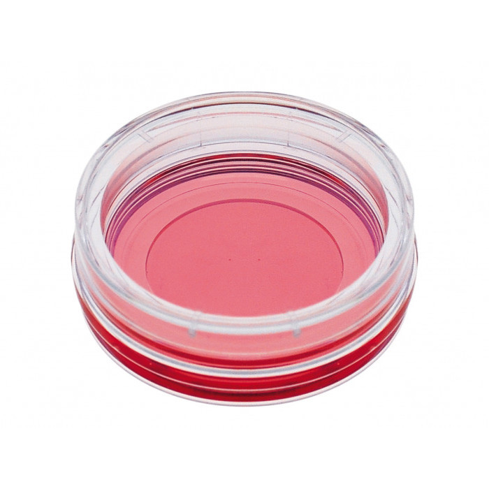
Glass Bottom Dish 35 mm
A 35 mm cell imaging dish with a glass bottom for everyday cell culture and fluorescence microscopy
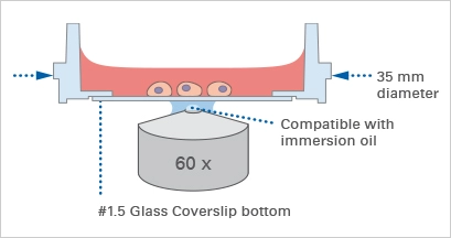
- Cost-effective cell culture dish with a glass coverslip bottom (#1.5)
- Excellent optical properties for high-resolution microscopy
- Very low autofluorescence
Applications
- Cultivation and high-resolution microscopy of cells
- Cost-effective cell imaging through a glass coverslip bottom
- Sensitive fluorescence analysis (e.g., FRET, FRAP, FLIM, and TIRF) possible, but we recommend the µ-Dish 35 mm, high Glass Bottom
- Immunofluorescence staining
- Widefield and confocal fluorescence microscopy of living and fixed cells
- Live cell imaging over extended time periods
- Transfection assays
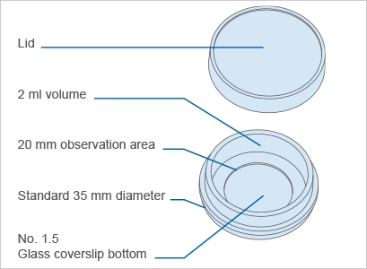
Specifications
| Ø Dish | 35 mm |
| Volume | 2 ml |
| Growth area | 3.14 cm² |
| Coating area using 400 µl | 3.7 cm² |
| Ø observation area | 20 mm |
| Height with / without lid | 11.7 mm / 9.9 mm |
| Bottom Glass | #1.5 glass coverslip, 170 µm (+20/-10 µm) |
Technical Features
- Standard format imaging dish with a 35 mm diameter for tissue culture
- Cover glass bottom made from D 263 M Schott glass with a thickness of 170 µm (- 10 µm / + 20 µm)
- May require coating to promote cell attachment
- Packed in sleeves with 10 pieces each
- 200 or 800 pieces per box
- Not compatible with DIC Lids for µ-Dishes
Find more information here
Other Products
-

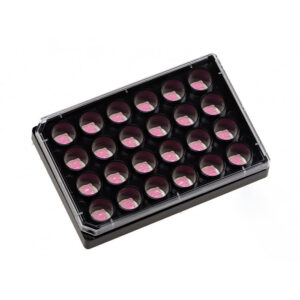
Culture-Insert 2 Well 24
Select options This product has multiple variants. The options may be chosen on the product page -
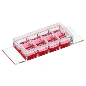
µ-Slide 8 Well high Bioinert
Select options This product has multiple variants. The options may be chosen on the product page -
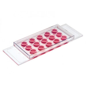
µ-Slide 15 Well 3D (formerly µ-Slide Angiogenesis)
Select options This product has multiple variants. The options may be chosen on the product page -
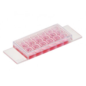
µ-Slide 18 Well
Select options This product has multiple variants. The options may be chosen on the product page