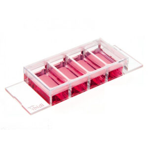
µ-Slide VI 0.4 Bioinert
A 6 channel μ-Slide with a non-adherent surface for flow experiments and fluorescence microscopy
- Superior to the standard ultra-low attachment (ULA) surfaces—stable, biologically inert surface for long-term experiments without any cell or biomolecule adhesion
- Excellent microscopic quality without any autofluorescence—100% surface passivation combined with the highest optical quality of the ibidi Polymer Coverslip
Applications
- Real-time imaging under either static or flow conditions
- Generation and long-term culture of 3D cell aggregates (e.g., spheroids and organoids) without any scaffold
- High resolution fluorescence microscopy of organoids, spheroids, embryoid bodies (EBs), and cells in suspension
- Background-free analysis of cell-cell interactions
- Prevention of stem cell differentiation due to attachment
- Culture of suspension cells in a permanently unattached state
- Self-assembly tumor spheroid formation assays
- 3D tumor spheroid models
Specifications
Bioinert Surface
| Bioinert surface thickness | 200 nm |
| Bioinert surface material | Polyol-based hydrogel |
| Protein coatings | Not possible |
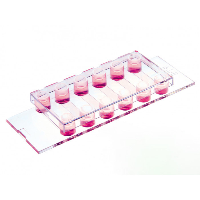
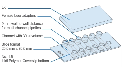
µ-Slide VI 0.4
| Outer dimensions (w x l) | 75.5 x 25.5 mm² |
| Adapters | Female Luer |
| Number of channels | 6 |
| Channel volume | 30 µl |
| Channel height | 0.4 mm |
| Channel length | 17 mm |
| Channel width | 3.8 mm |
| Volume per reservoir | 60 µl |
| Growth area | 0.6 cm² per channel |
| Coating area using 30 µl | 1.2 cm² per channel |
| Bottom: ibidi Polymer Coverslip with Bioinert surface | |
Technical Features
- Channel µ-Slide with 6 independent channels and a non-removable polymer coverslip-bottom
- µ-Slide VI 0.4 with a non-adherent, passivated Bioinert surface
- Bioinert surface with long term passivation—superior to standard ULA surfaces:
- Ready-to-use without prior surface treatment or preparation
- No adsorption, coating, or binding of proteins, antibodies, enzymes, and other biomolecules
- Non-cytotoxic, biologically inert, and non-degradable
- Easy connection to existing tubes and pumps via female Luer adapters
- Imaging µ-Slide with excellent optical quality for high-end microscopy
- Compatible with staining and fixation solutions
- Biocompatible polymer material—no glue, no leaking
Other Products
-

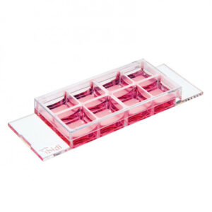
µ-Slide 18 Well high Glass Bottom
Select options This product has multiple variants. The options may be chosen on the product page -
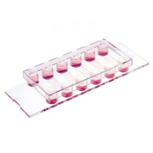
µ-Slide 4 Well Glass Bottom
Select options This product has multiple variants. The options may be chosen on the product page -
µ-Slide VI 0.4
Select options This product has multiple variants. The options may be chosen on the product page -
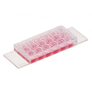
µ-Slide 8 Well
Select options This product has multiple variants. The options may be chosen on the product page