ibidi Photo Contest – Call for Entries: 2027 ibidi Calendar


Bring Your Best Shot: 2027 ibidi Calendar

Your next great microscopy image deserves to be seen. The 2027 ibidi Photo Contest is officially open for submissions!

If you have a microscopy image that made you stop and look twice, this is your moment to share it. 12 winners will have their images featured in the 2027 ibidi calendar and will receive 3 free copies. Plus, the winners' images will also be showcased on the ibidi website and social media channels.

Take a moment to explore the winning images from the 2026 ibidi Calendar, and don’t forget to visit our ibidi CellArt Gallery for even more creative insights.

It’s time to showcase your microscopy images and research! Be a part of making the ibidi 2027 calendar outstanding!

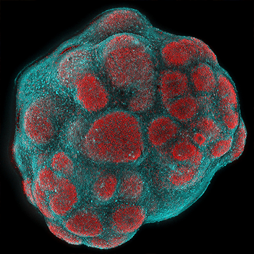
One of 2026’s Captivating Images

Stephanie Le
Heinrich Heine University, Düsseldorf, Germany

Young cortical organoids imaged in a μ-Slide 8 Well high Glass Bottom show SOX2-positive progenitor cells (red) clustering in proliferative zones, while MAP2-positive neurons (cyan) extend early processes. This image reveals the spatial organization and early differentiation dynamics of the developing human cortex in vitro. Imaging was performed on a Zeiss Axio Observer microscope with a 5x objective.

https://campaign-image.eu/zohocampaigns/14215000045874218_zc_v32_1621500337381_ibidi_calendar_image_2021.jpg
   

Deadline for submission is June 30, 2026. The winners will be selected in September 2026.


SVI Huygens 26.04 Released with Cellpose 3D! And Live Webinar on April 28th, May 12th

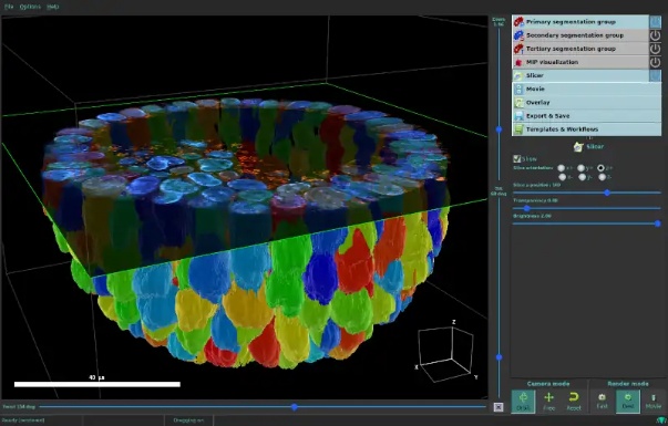
Huygens 26.04 is released with Cellpose 3D, newROI Options, Bioformats compatible Huygens HDF5, faster Stitcher, Object Analyzer features, and more!

What’s in 26.04 – Live Webinar April 28th

We’ll show Cellpose 3D integration, Pyramid HDF5 compatibility with Bioformats/BDV, free camera movement around and through 3D objects, coloc analysis per ROI element, drastic speed improvement in stitching and vignetting correction, and more…

SMLM analysis – Live Webinar May 12th

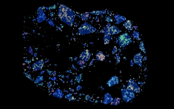
Fast, accurate, and easy SMLM reconstruction and analysis with Huygens Localizer – featuring Nucleosome cluster analysis.

NanoInsights in Chinese Society for Cell Biology (中国细胞生物学学会 2026)

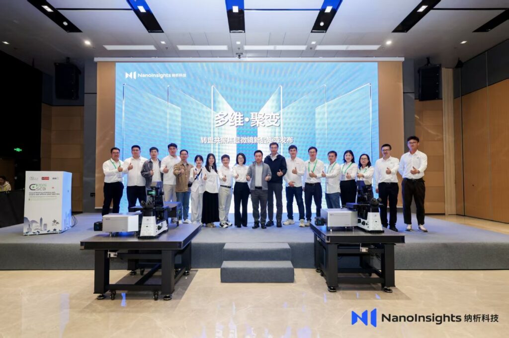
Our Principal NanoInsights participated in the 2026 National Academic Conference of the Chinese Society for Cell Biology (中国细胞生物学学会2026年全国学术大会) and announcing the joined-force with Pellucid Optics and their new product – SuperSpin and PanoSpin Label-free imaging system

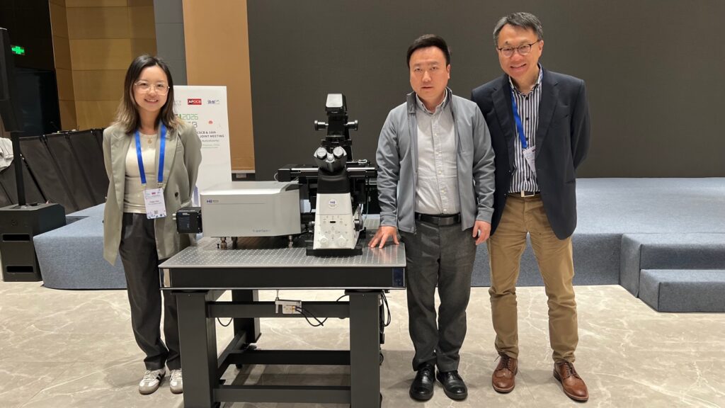
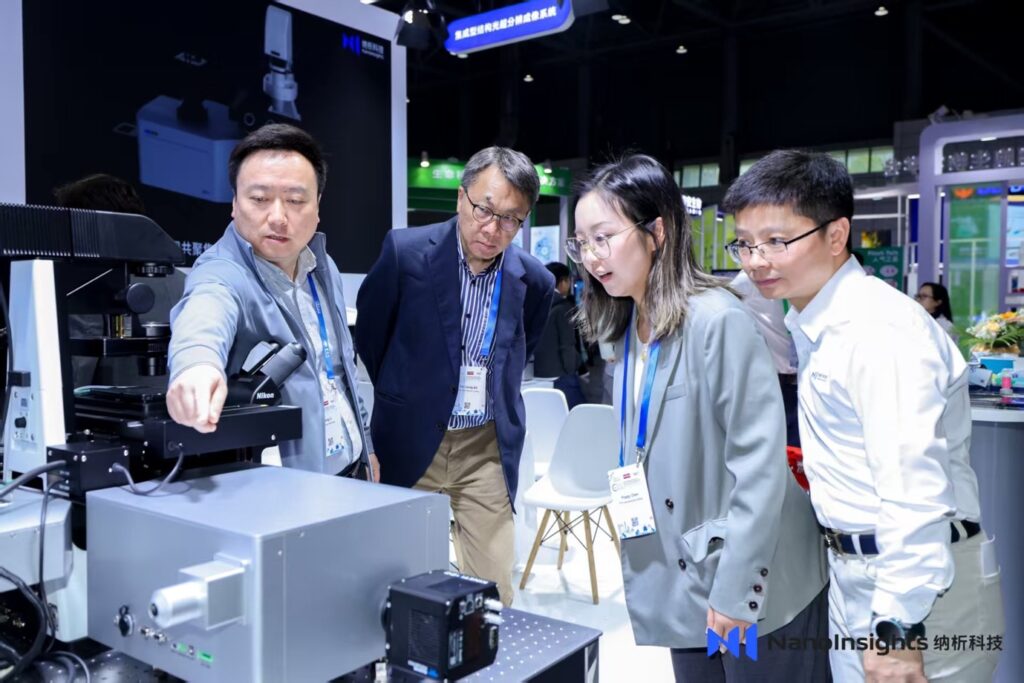
From left: Dr. Poppy Chen (PTC), Professor Dong Li (Founder of NanoInsights) and Dr. CL So (PTC)
Professor Li (Left)explaining the unique superresolution functionality in “SuperSpin” to Dr. Chen (second right) and Dr. So (second left) in NanoInsights’ Booth. Mr. Guo, the sales manager of the NanoInsights (right).

Abberior in Chinese Society for Cell Biology (中国细胞生物学学会 2026)

Our Principal Abberior Instruments GmbH participated in the 2026 National Academic Conference of the Chinese Society for Cell Biology (中国细胞生物学学会2026年全国学术大会).

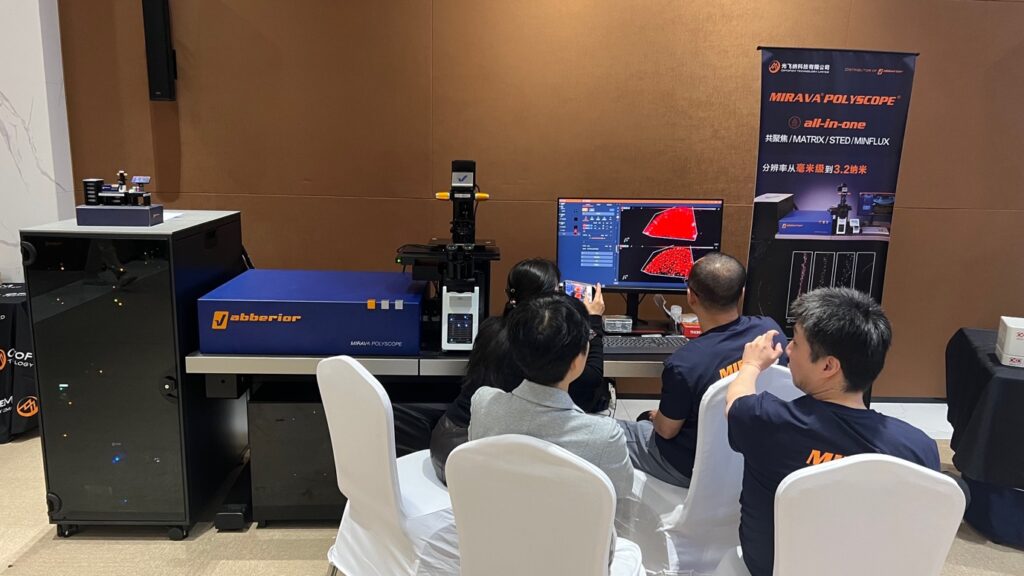
The MIRAVA Polyscope was demonstrated in the show room with fully functionalities, including the 2D MINFLUX nanoscale imaging, 3D STED, MATRIX array detection, RAYSHAPE and TRUESHARP imaging.
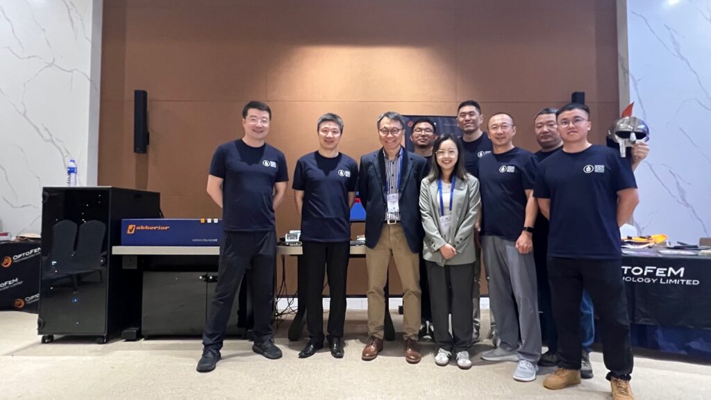
The Distributors of Abberior Optofem (China), PTC (Hong Kong) and the Abberior team (China).

PTC is the Official Distributor of Pellucid Optics (澄觀光學)

PTC is now the Official Distributor of Pellucid Optics (澄觀光學科技有限公司) in Hong Kong and Macau.

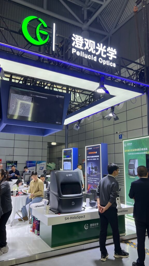
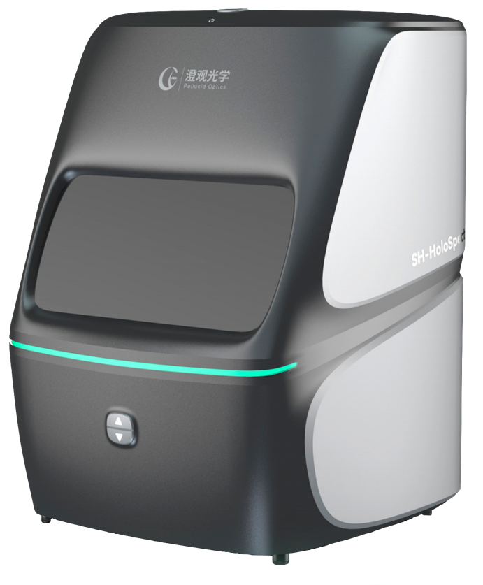
The latest Label-free 3D Volumetric Imaging System: SH-HoloSpect was first displayed in the 2026 National Academic Conference of the Chinese Society for Cell Biology (中国细胞生物学学会2026年全国学术大会).

Pellucid Optics is specialized in Label-Free-Superrsolution Imaging Technology. Their unique Phase Gradient Tomography (PGT) allows layer scanning, up to 140nm lateral and 350nm axial resolution, to render a 3D Volumetric Image.

SH-HoloSpect

  1. Label-free subcellular imaging
    Without the need for complex labeling or staining procedures, subcellular structures such as granules, nuclei, and lipid droplets can be accurately identified based on intrinsic differences in refractive index.
  2. Advanced PGT imaging capabilities
    The new generation of PGT technology supports both 2D and 3D sample imaging. It is compatible with multiple imaging modes, including wide-field fluorescence, spinning-disk confocal multi-module imaging, and high-throughput imaging workflows.
  3. Comprehensive data analysis software
    The included software enables automated collection and analysis of key biophysical parameters, including refractive index, dry mass, velocity, and cellular morphology.