PTC is granted the only Official Distributor and Certified Gold Partner of Scientific VolumeImaging software in Hong Kong and Macau.
The Huygens Software is named after Christiaan Huygens, who studied and described the diffraction of light waves. The Software includes many image processing options, for brightfield and all types of fluorescence Microscope images. With Huygens it is possible to perform image deconvolution and restoration, interactive analysis and volume visualization in 2D-4D, multi-channel and time. Additional Server and GPU-based Options ensure maximization of hardware performance.
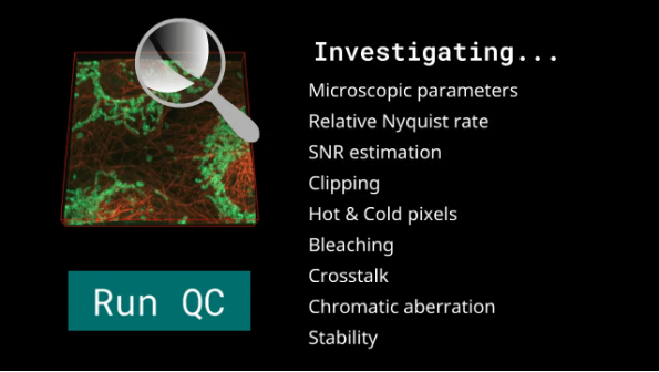
The Image Quality Control (QC) tool can analyse images and bead data thoroughly, alerting users any potential problems, such as microscopic parameters, crosstalk, chromatic aberration, stability and bleaching issues. Each detected issue can be directly verified and fixed through the QC tool.
Better yet, a free online version – with limited features – is available here for trial.
Huygens Software
Huygens offers deconvolution for postprocessing of raw data from different kinds of microscopy, like confocal, widefield, Multi-photon, Spinning-disk, STED, Array-detector, Light-sheet, Rescan, Visitech, Aurox, 4-pi, etc., and various metadata is supported including Leica LIF; Zeiss CZI, LSM, ZVI; Nikon ND2, Deltavion DV, OME TIFF, Abberior OBF, MSR; and many more! The theoretical or measured PSF is fed into the most advanced MLE algorithms that currently exist to restore the image and to effectively correct blur, noise and background. Note that the Huygens MLE algorithms conserve the intensities, so your deconvolution results will still be quantifiable, and even more so, the deconvolved results will be much easier to analyze compared to the raw data.
Restore the information lost in microscopy imaging
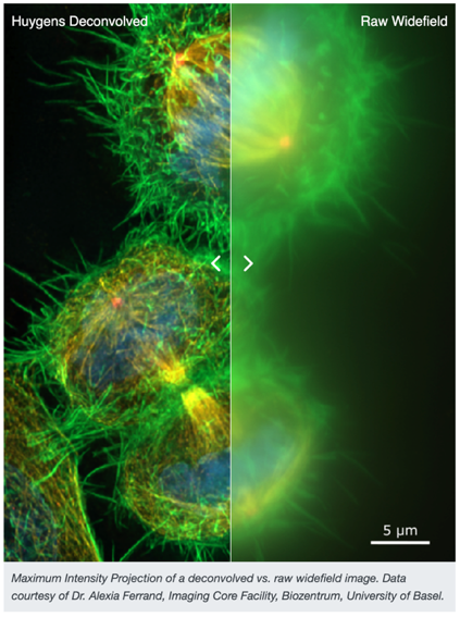
Maximum Intensity Projection of a deconvolved vs. raw widefield image. Data courtesy of Dr. Alexia Ferrand, Imaging Core Facility, Biozentrum, University of Basel.
Correct XYZshifts, rotations, scaling and radical differences between channels
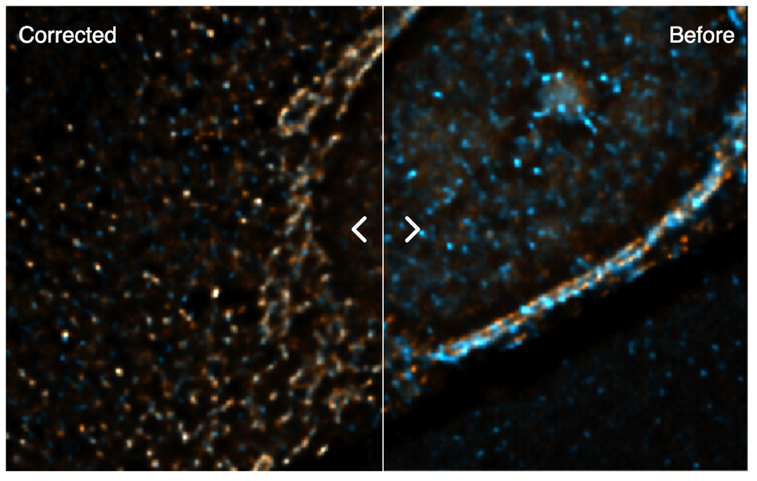
HeLa cells stained with two different fluorophores. This image shows a shift between the two different channels which has been corrected with Huygens Chromatic Aberration Corrector. Raw image data was provided by Dr. Yury Belyaev, ALMF, EMBL Heidelberg, Germany
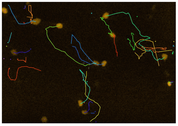
Study 3D motion of cells and small particles in time series
FUCCI cells tracked for 50 seconds with Huygens Object Tracker. Courtesy of Dr. Richard Wubbolts, Center for Cell Imaging, Faculty of Veterinary Medicine, University of Utrecht, The Netherlands.
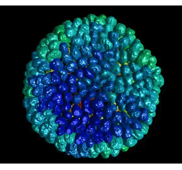
Complete organoid visualized using Huygens Surface Renderer. The original confocal image of 493 Z-planes of an organoid stained for nuclei was first deconvolved and then rendered with Huygens. Courtesy of Dr. Rolando Berlinguer Palmini, BioImaging Unit, Newcastle University, UK
Obtain projections of the highest possible quality
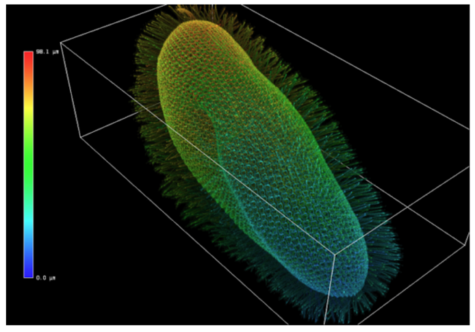
The MIP renderer in color mode depth-coding showing a Paramecium. Regions of the image close to the viewpoint appear blue whereas region far away appear red. Image courtesy of A. Aubusson-Fleury CNRS, Gif sur Yvette, Paris.
Would like to obtain more info and learn more of its applications?
PTC is granted the only Official Distributor and Certified Gold Partner of Scientific VolumeImaging software in Hong Kong and Macau.
The Huygens Software is named after Christiaan Huygens, who studied and described the diffraction of light waves. The Software includes many image processing options, for brightfield and all types of fluorescence Microscope images. With Huygens it is possible to perform image deconvolution and restoration, interactive analysis and volume visualization in 2D-4D, multi-channel and time. Additional Server and GPU-based Options ensure maximization of hardware performance.
The Image Quality Control (QC) tool can analyse images and bead data thoroughly, alerting users any potential problems, such as microscopic parameters, crosstalk, chromatic aberration, stability and bleaching issues. Each detected issue can be directly verified and fixed through the QC tool.
Better yet, a free online version – with limited features – is available here for trial.
Huygens Software
Huygens offers deconvolution for postprocessing of raw data from different kinds of microscopy, like confocal, widefield, Multi-photon, Spinning-disk, STED, Array-detector, Light-sheet, Rescan, Visitech, Aurox, 4-pi, etc., and various metadata is supported including Leica LIF; Zeiss CZI, LSM, ZVI; Nikon ND2, Deltavion DV, OME TIFF, Abberior OBF, MSR; and many more! The theoretical or measured PSF is fed into the most advanced MLE algorithms that currently exist to restore the image and to effectively correct blur, noise and background. Note that the Huygens MLE algorithms conserve the intensities, so your deconvolution results will still be quantifiable, and even more so, the deconvolved results will be much easier to analyze compared to the raw data.
Restore the information lost in microscopy imaging
Maximum Intensity Projection of a deconvolved vs. raw widefield image. Data courtesy of Dr. Alexia Ferrand, Imaging Core Facility, Biozentrum, University of Basel.
Correct XYZshifts, rotations, scaling and radical differences between channels
HeLa cells stained with two different fluorophores. This image shows a shift between the two different channels which has been corrected with Huygens Chromatic Aberration Corrector. Raw image data was provided by Dr. Yury Belyaev, ALMF, EMBL Heidelberg, Germany
Study 3D motion of cells and small particles in time series
FUCCI cells tracked for 50 seconds with Huygens Object Tracker. Courtesy of Dr. Richard Wubbolts, Center for Cell Imaging, Faculty of Veterinary Medicine, University of Utrecht, The Netherlands.
Complete organoid visualized using Huygens Surface Renderer. The original confocal image of 493 Z-planes of an organoid stained for nuclei was first deconvolved and then rendered with Huygens. Courtesy of Dr. Rolando Berlinguer Palmini, BioImaging Unit, Newcastle University, UK
Obtain projections of the highest possible quality
The MIP renderer in color mode depth-coding showing a Paramecium. Regions of the image close to the viewpoint appear blue whereas region far away appear red. Image courtesy of A. Aubusson-Fleury CNRS, Gif sur Yvette, Paris.
Would like to obtain more info and learn more of its applications?