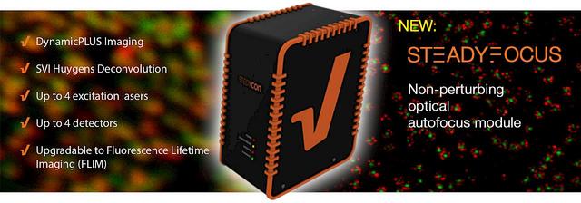
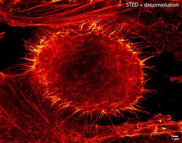
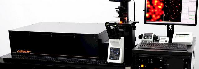
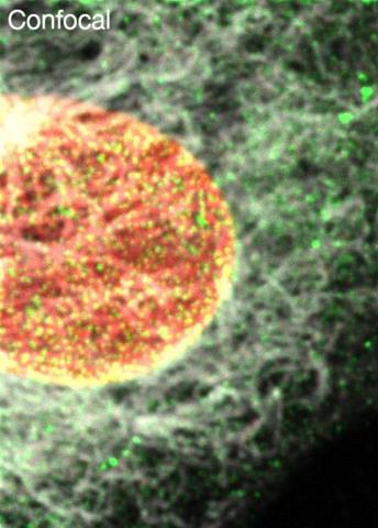
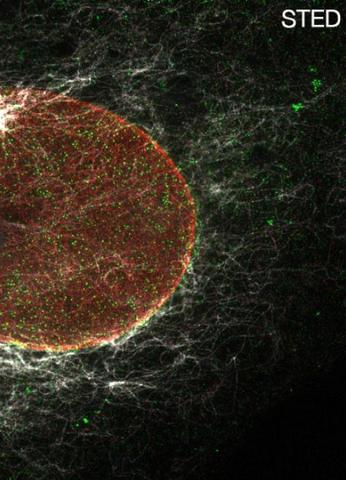
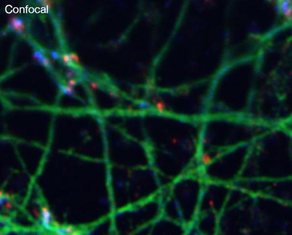
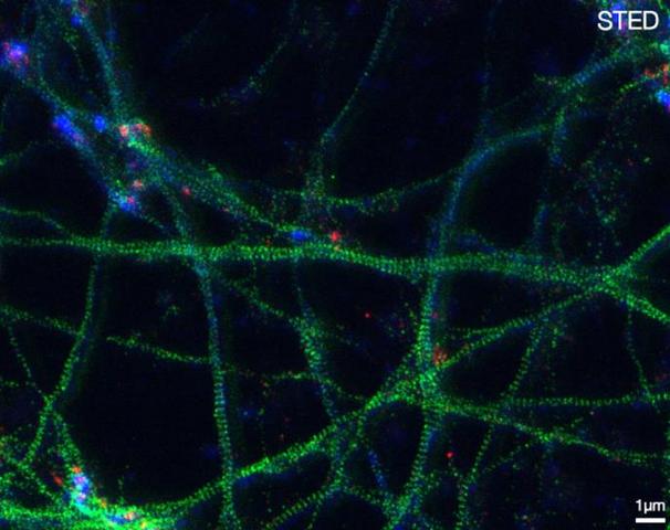
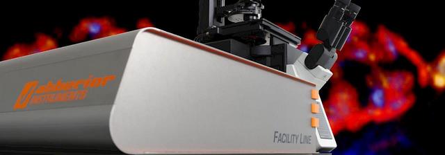
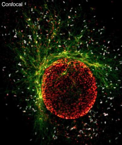
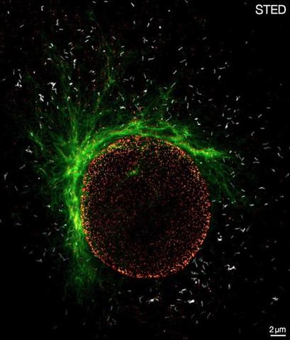
Abberior Instruments is known for extraordinary customization ability of its flexible STED microscope platform. Abberior has developed the ultimate compact STED and confocal microscope, called STEDYCON.
STEDYCON is a completely new class of nanoscope. It converts your conventional epifluorescence microscope into a versatile four-color confocal (405nm, 488nm, 561nm, 640nm) and STED (775nm) system! At the same time, it is incredibly compact and can be used by anyone!